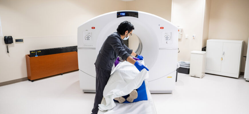
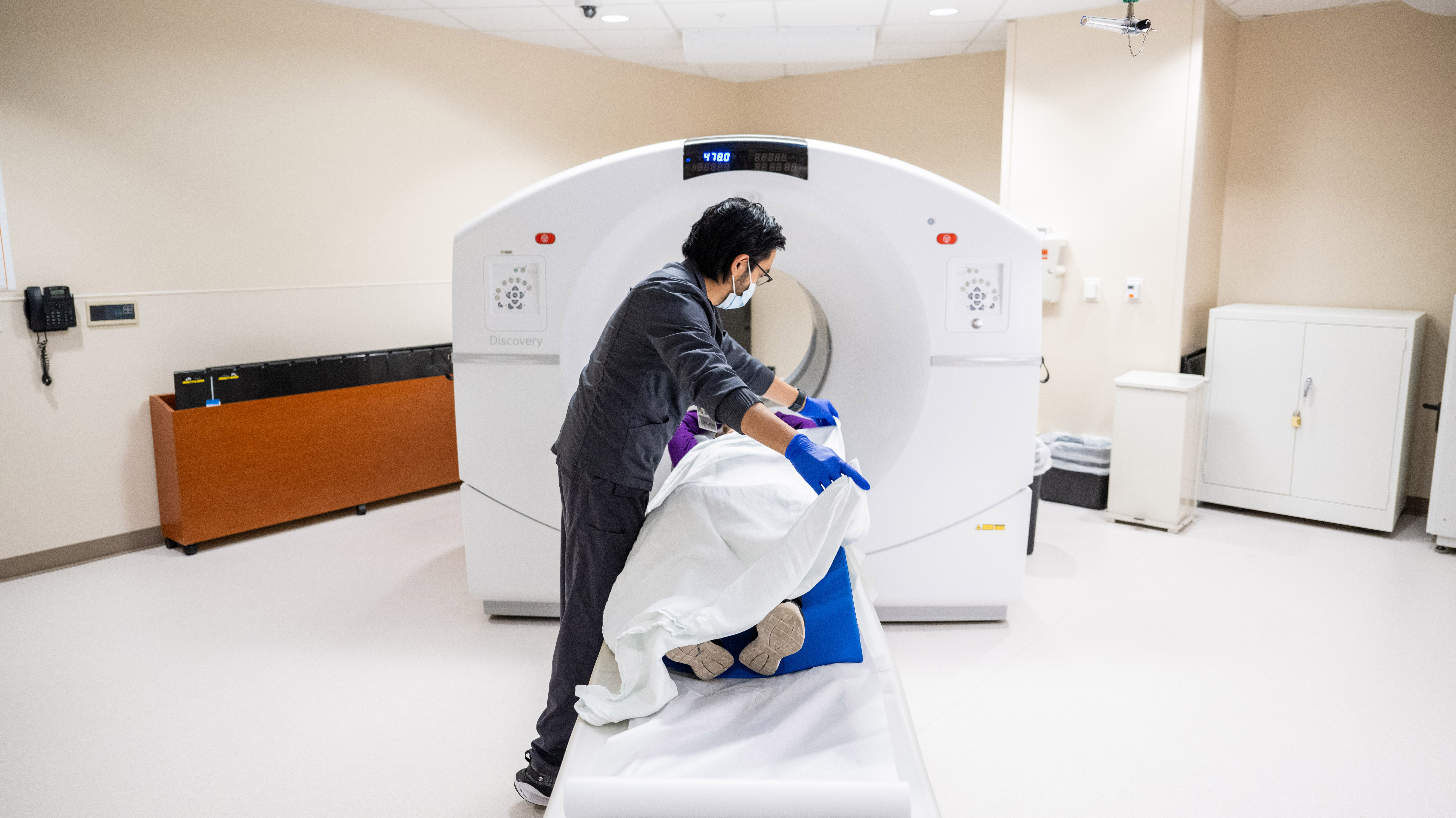
A doctor orders a PET scan to detect cancer, check if it has spread, or evaluate heart and brain problems by showing how active tissues are.
What it shows
A PET (positron emission tomography) scan uses a radioactive tracer, usually FDG (a sugar), to show how tissues use energy. Active areas, such as tumors, light up.
Common reasons
- Detect cancer and check if it has spread.
- Check treatment response.
- Evaluate heart blood flow and damage.
- Evaluate brain disorders such as Alzheimer’s and epilepsy.
Often paired
PET/CT combines function and anatomy.
Prep
Fast 4 to 6 hours. Scan takes 30 to 60 minutes.
Radiation
Similar to a few years of natural background exposure.
How the scan works in numbers
PET (positron emission tomography) uses a radiotracer, most often fluorodeoxyglucose (FDG), a glucose analog labeled with fluorine-18, which has a half-life of about 110 minutes. Cells that use more glucose, such as many tumors, active inflammation and brain regions at work, take up more tracer. A typical whole-body scan takes 20 to 30 minutes after an uptake period of about 60 minutes.
- Standard uses include staging and restaging cancers (lymphoma, lung, melanoma, colorectal), checking treatment response, and finding recurrence.
- In neurology, amyloid and FDG PET help evaluate Alzheimer disease and epilepsy foci.
- In cardiology, PET measures blood flow and viable heart muscle.
- Most scans are combined with CT (PET/CT) so uptake can be matched to anatomy.

Preparation and safety
Patients usually fast for 4 to 6 hours and avoid strenuous exercise for 24 hours. People with diabetes need instructions on blood sugar, since high glucose lowers FDG uptake. The effective radiation dose of an FDG PET/CT is roughly 7 to 14 mSv, depending on the CT protocol, compared with about 3 mSv average annual natural background in the US. Pregnant or breastfeeding patients should tell staff, and some pause breastfeeding for a few hours.
Technology and dose
A PET scan uses a radioactive tracer, most commonly fluorodeoxyglucose (FDG), a sugar analogue that accumulates in cells with high metabolism such as cancers. The tracer has a half life of about 110 minutes. A typical whole body PET scan involves an effective radiation dose of about 7 to 14 millisieverts, and PET/CT is higher. Scanning begins about 60 minutes after injection and lasts 20 to 30 minutes.
Preparation
Patients usually fast for 4 to 6 hours, avoid strenuous exercise the day before and keep blood sugar controlled. Diabetics need specific instructions. Tell the team if you are pregnant or breastfeeding. Drink water afterward to help clear the tracer. PET is used for cancer staging, heart disease and brain conditions such as Alzheimer’s disease.
Dose, timing and accuracy
PET scans detect metabolic activity, not just structure. Typical injected FDG dose is about 370 MBq (10 mCi). Studies show PET/CT changes the cancer stage in roughly 10 to 30 percent of patients compared with CT alone. A scan takes about 2 hours in total, including uptake time.

Preparation and limits
Patients avoid sugar and fasting for 4 to 6 hours; for diabetic patients, glucose levels should be below about 200 mg/dL. Brain PET is used in Alzheimer’s evaluation with amyloid tracers. False positives happen with infection and inflammation. The radioactivity decays quickly, but hugging infants or pregnant women is limited for several hours.
Based on data from the Society of Nuclear Medicine and Molecular Imaging and the American College of Radiology